
Talk:Microscopy
| This article is rated B-class on Wikipedia's content assessment scale. It is of interest to the following WikiProjects: | |||||||||||||||||||||||||||||||||||||||||
| |||||||||||||||||||||||||||||||||||||||||
.jpg)


Sub-diffraction limit techniques[edit]
Currently the sub-diffraction limit section is a mess, it is badly written, quickly changing and far more complex than most of the rest of this article; would it make sense to split it to a separate article? - Zephyris Talk 10:13, 27 July 2009 (UTC)
STED, SIM, PALM, STORM, FPALM[edit]
Hi,
the section on super-resolution fluorescence microscopy (i.e. techniques like STED, SIM, PALM, STORM, FPALM,...) is really outdated. That field has developed rapidly over the last two years. Maybe someone could provide an update. —Preceding unsigned comment added by 134.76.215.201 (talk) 22:10, 20 November 2008 (UTC)
A polite request...[edit]
I am doing a major overhaul of this page over the course of the next week or two, and its not finished yet! so please don't revert all of my changes because it seems a bit unstructured, it will get better :D Zephyris 21:12, 6 August 2006 (UTC)
+++ Question +++ Are Particle Colliders and Atom Smashers not some sort of microscopes? If so, could you include some appropriate links to relevant articles on that? William vroman
- yes, in some respects. my feeling is that microscopy refers to any mechanism which 'directly' images a target, which does apply to some particle collider experiments. I think that some experiments, such as electron(?) scattering to reveal neutron/proton quark structures, would be appropriate here, and a short mention in the introduction and a short section (as for scanning probe microscopes) would be suitable. - Zephyris Talk 01:38, 18 November 2006 (UTC). Sorry if this doesnt make much sense - wine at work!
- I'd draw the line at what you can find in good sources. If you find a book that refers to these things as microscopy, go for it. Dicklyon 06:31, 18 November 2006 (UTC)
Action[edit]
I have read through the talk here and at microscope to try and get something done about the future of these articles. It seems that people agree on what should be done, it just needs doing!
Here is an outline of what I believe a sensible solution is for the microscopy, microscope and all other related pages:
- Microscope and microscopy should become portal style pages, with summary articles and links to pages on the individual types of microscopy (optical, electron, etc.) and pages on the physical principles of basic microscopes (ie. optics, resolution, electron optics, etc.)
- Microscope should be written from a physical viewpoint, ie. the physics and history of microscopes, as microscopes are the actual instrument. Microscopy should be written from a more practical viewpoint, ie. the usage and reasons for usage of the different techniques.
- Optical microscope needs its own page, similar to electron microscope. Relevant information on individual optical microscopy instruments and techniques need to be moved to this page.
- Each individual microscopy technique and microscope type (eg. phase contrast, scanning electron, etc.) needs its own page, no matter how short - it is better to have a stub for expansion than a long and confusing parent article.
Finally and most importantly:
- microscope and microscopy should be kept short and simple. They are introductory pages to what is a very wide and in depth region of science. Detail should be confined to more focussed articles.
You have a week to make your comments, and, unless there are any major complaints, im going to get started! Zephyris 20:42, 30 July 2006 (UTC)
- Comment: nothing you propose seems crazy. Why wait? I cannot imagine that anyone will revert you if you make these reasonable changes. Be bold in updating pages. --JWSchmidt 22:05, 30 July 2006 (UTC)
- Thanks for the confidence boost :D, and i have now let myself loose on the articles, bwahahaha! Zephyris 21:12, 6 August 2006 (UTC)
Take a look at microscope and consider the relationship between these two articles. adam
- I think that has been done - Marshman 03:51, 30 Mar 2005 (UTC)
This article needs urgent revision. Porcher 23:14, 29 Mar 2005 (UTC)
- and it still needs urgent revision. Karol 22:11, 30 December 2005 (UTC)
Agreed that this needs substantial improvement, but I would oppose any merge with Microscope. A microscope is the instrument or the tool with which work is done. Microscopy describes the techniques in which a microscope is used. It would be like merging Automobile and Driving
Velela 16:59, 21 February 2006 (UTC)
The inventor of DIC seems to be called Georges Nomarski. Gogowitsch 09:55, 4 May 2006 (UTC)
--- Do you consider deconvolution as another kind of microscopy? I guess that deconvolution should be explained in post-acquisition or image restoration techniques, as a complement for e.g. fluorescence microscopy.... Default007 14:02, 26 July 2006 (UTC)
Link suggestion[edit]
Is this link relevant for this webpage (page-owner, self-promotion?)? Pvosta 11:33, 12 September 2006 (UTC)
Biomedical imaging - information about/for microscopy http://ourworld.compuserve.com/homepages/pvosta/pcrimg.htm
Is this link relevant for this webpage (page-owner, self-promotion?)? Pvosta 11:33, 12 September 2006 (UTC)
Nyquist Sampling in Digital Microscopy - sampling theorem: http://ourworld.compuserve.com/homepages/pvosta/pcrnyq.htm
Polarised light microscopy and Multiphoton microscopy??[edit]
Shouldn't these be included? Rotational 05:41, 7 November 2007 (UTC)
Specular microscopy[edit]
Is there a page on specular microscopy already, or it is needed to be created? --CopperKettle (talk) 21:47, 27 September 2008 (UTC)
This is a new article if anyone wants to have a look. ChildofMidnight (talk) 23:05, 3 August 2009 (UTC)
Image suggestion[edit]
I think this might be of use for illustrating the differences between light and electron microscopy. It's currently used on several butterfly-related articles so if you need to make significant changes that might negatively affect the placement in those articles, I recommend creating a fork:
Photographic and light microscopic images 

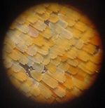
Zoomed-out view of an Aglais io. Closeup of the scales of the same specimen. High magnification of the coloured scales (probably a different species). Electron microscopic images 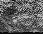


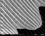
A patch of wing Scales close up A single scale Microstructure of a scale Magnification Approx. ×50 Approx. ×200 ×1000 ×5000


Papa Lima Whiskey (talk) 11:33, 1 May 2010 (UTC)
The set is interesting, but the individual images have quality issues, making the resultant set of less-than-encyclopedic quality for illustrating a microscopy article.
The light micrographs do not have scale markers on them, and the last one is out of focus in all planes. The 1000X electron micrograph is also out of focus everywhere in the image. Insect images taken on the SEM should have remarkable depth of field, showing many object planes in focus in a single micrograph, and thus illustrating one of the characteristics of the instrument. All of the scanning electron micrographs also have a lot of charging.
While the image set may work well for illustrating biological concepts, as micrographs they can only be used individually to illustrate improper focus, poor depth of field, or improper grounding or non-coating interfering with the scanning image formation. --KMLP (talk) 00:58, 11 October 2010 (UTC)
Lead section[edit]
This would ultimately make a great featured article, but still needs a lot of work, as with any article this large in scope of topic. I am removing part of the lead, not because the information does not belong in the article or even in the lead, but to focus initially on the overall readability and usability of the article for the general reader of an encyclopedia. This sentence can be incorporated elsewhere for now, or back to the lead as editors develop it:
- "along with many others including materials science and forensic engineering where microstructure and surface details such as trace evidence are important for understanding properties or the causes of accidents."
While microscopy is fundamental for materials science, it does not necessarily trump its importance in the biological or physical sciences in general. An additional paragraph discussing the importance of microscopy as a technique in the biological and physical sciences plus materials science in particular could appropriately develop the lead section with this sentence. I did leave "revolutionized biology," in because microscopy did that, while it contributed greatly to the revolution of other areas of science. --KMLP (talk) 17:34, 12 October 2010 (UTC)
Atomic de Broglie - does it exists?[edit]
As far as I know there is no Atomic de Broglie microscope anywhere on Earth. I strongly believe this entry should be deleted. There is a separate article on the subject, and it is sufficient. Mentioning Atomic de Broglie "microscope" among working techniques is misleading at least.
Limitations[edit]
What happened in this sentence: "Staining may also introduce artifacts, apparent structural details that are caused by the processing of the specimen ame extent by specific microscopy techniques that can non-invasively increase the contrast of the image."? I don't know enough about microscopy to correct this sentence, but it really should be revised. 129.206.24.62 (talk) 13:08, 12 June 2014 (UTC)
Request some metrics for comparing microscopy techniques, and a table comparing them[edit]
I'd like to start by thanking those who know something about microscopes for writing into this page for those of us who know little!
I looked this up this page after getting an ad for "affordable atomic force microscopes" because I had no clue about them. Sadly, after reading the article, I still don't know how on what bases I would choose a microscope if I had the need. Part of this is needing metrics. Here is my beginning of a list of the metrics, based on what I (as a non-expert) would anticipate being important:
- finest demonstrated resolution of the microscopy technique in each dimension. Are width and length equal for all techniques? I doubt it. Which techniques can measure depth?
- dimensions and units of measurement - optical microscopy, of course, allows many dimensions aligning with color (subject to diffraction limits etc); is electron microscopy a bunch of bits, or perhaps 8-bit values?
- how much damage the measuring technique does to the sample - atomic-force microscopes seem to scrape the surface; dyes change the chemistry...
- ability of a technique to resolve multiple layers in a sample
- capture pattern, e.g. scanning
- ability to image live specimens
- scanning speed in area/time - square-mm per second I suppose
- restrictions on size or other characteristics of the sample
With that done, I would propose setting up a table listing microscopy techniques and these dimensions, obviously to allow the reader to sort by one (or more?) dimensions of interest.
Schemaczar (talk) 19:23, 8 June 2017 (UTC)
Reflected-light microscopy[edit]
I can't find any useful discussion of reflected-light optical microscopy. While little used in biology for very small targets, it seems very important in other sciences (industry, geology, etc.) and even for botanics and entomolog.--Jorge Stolfi (talk) 19:13, 12 June 2017 (UTC)
A Commons file used on this page or its Wikidata item has been nominated for deletion[edit]
The following Wikimedia Commons file used on this page or its Wikidata item has been nominated for deletion:
Participate in the deletion discussion at the nomination page. —Community Tech bot (talk) 04:17, 5 January 2022 (UTC)
- B-Class Microbiology articles
- Top-importance Microbiology articles
- WikiProject Microbiology articles
- B-Class Molecular Biology articles
- Unknown-importance Molecular Biology articles
- B-Class MCB articles
- Top-importance MCB articles
- WikiProject Molecular and Cellular Biology articles
- All WikiProject Molecular Biology articles
- B-Class medicine articles
- Mid-importance medicine articles
- All WikiProject Medicine articles
- B-Class Technology articles
- WikiProject Technology articles